Cholesterol granuloma

Low confidence
High confidence
- Age: 64
- Sex: Female
- Modality: MRI
- Region: Brain
- Country: N/A
- State: N/A
- City: N/A
- Diagnosis: Cholesterol granuloma
🧠 AI Suggestion
1) Findings:
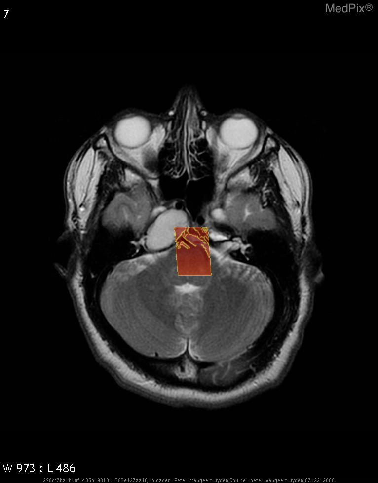
- Axial MRI brain shows a large, well-defined, expansile lesion centered in the petrous apex region on the image-left (patient-right).
- Lesion demonstrates homogeneously high signal intensity on T2-weighted sequence, suggesting fluid or blood product content.
- The mass appears to cause remodeling and expansion of the petrous temporal bone on the image-left (patient-right).
- No definite internal septations or solid enhancing nodules are visible on this T2 image.
- Adjacent structures such as the cerebellum and pons are mildly compressed but not invaded.
- The middle ear and mastoid on the image-left (patient-right) appear partially effaced.
- Optic apparatus and contralateral (image-right, patient-left) structures are normal.
- No diffusion restriction or perilesional edema is observed on this sequence (though confirmation requires DWI).
2) Most likely diagnosis and why:
Cholesterol granuloma of the petrous apex (image-left = patient-right). This diagnosis is favored due to the well-circumscribed expansile lesion in the petrous apex with homogeneous T2 hyperintensity and smooth bony expansion, typical for a cholesterol granuloma containing blood degradation products. No solid component or irregular enhancement pattern suggests a cystic, benign process rather than neoplastic. Context consistency: Consistent — matches the provided context of petrous apex cholesterol granuloma in a 64-year-old female. Confidence: 90%
3) Next best diagnostic step:
Dedicated high-resolution T1-weighted pre- and post-contrast MRI sequences of the petrous apex would confirm the absence of enhancement and show high intrinsic T1 signal from cholesterol and methemoglobin. Temporal bone CT can also be done to detail bony expansion and thinning. These steps help confirm the diagnosis and exclude cholesteatoma or other petrous apex cystic lesions.
4) Key differential or confirmatory test:
CT of the temporal bone is the key complementary test. Cholesterol granulomas show smooth expansion with cortical thinning, while cholesteatomas often cause bone erosion without significant expansion. MRI with diffusion-weighted imaging may also help as cholesteatomas restrict diffusion, unlike cholesterol granulomas.
5) Possible treatment or management:
** Management depends on the size, symptoms, and effect on adjacent cranial nerves. Asymptomatic lesions may be observed with serial MRI. Symptomatic lesions (e.g., hearing loss, cranial nerve deficits) may require surgical drainage or marsupialization via transsphenoidal or infralabyrinthine approach. The goal is to establish permanent drainage and prevent recurrence.
📑 Guidelines Summary (uploaded diagnosis) — Cholesterol granuloma
Imaging evaluation of suspected or known brain tumors focuses on optimal modality choice, treatment planning, and detection of complications or recurrence.
- New neurologic deficit, seizures, or cognitive change: MRI brain with and without contrast is first-line.
- CT head without contrast is appropriate for emergent assessment when MRI is unavailable or contraindicated.
- Postoperative or post-radiotherapy surveillance: MRI with and without contrast every 2–6 months in the first years.
- Suspected metastases: MRI with and without contrast preferred; consider contrast-enhanced CT if MRI unavailable.
- In acutely deteriorating patient with known tumor, non-contrast CT quickly assesses hemorrhage or herniation risk.
- Suspected vascular tumor component or hemorrhagic lesion: MRI with susceptibility or CTA for vessel involvement.
- Do not image for stable low-grade tumor without new or worsening symptoms outside scheduled follow-up.
- Red flags: rapidly progressive headache, focal deficit, seizure pattern change—urgent MRI with contrast indicated.
- Pitfalls include misinterpreting radiation necrosis or postoperative enhancement as tumor recurrence without advanced MRI sequences.
- Functional MRI and diffusion tensor imaging guide surgical planning in eloquent cortex involvement.
- Spectroscopy or perfusion MRI helpful in differentiating tumor progression from treatment-induced changes when conventional MRI equivocal.
🤖 Guidelines Summary (AI diagnosis) — Liver Lesion — Initial Characterization
Guidelines summarize optimal imaging choices for evaluating incidentally detected or suspected liver lesions based on clinical context and imaging features.
- In low-risk patients with ultrasound-detected lesion, multiphasic contrast-enhanced CT or MRI is first-line for characterization.
- For indeterminate lesions on prior CT, MRI with hepatobiliary contrast provides highest diagnostic accuracy.
- Non-contrast CT alone is inadequate for lesion characterization due to limited tissue differentiation.
- In patients with chronic liver disease or cirrhosis, multiphasic MRI or CT evaluates for hepatocellular carcinoma following LI-RADS criteria.
- Use contrast-enhanced ultrasound as alternative when MRI or CT contrast is contraindicated.
- Do not image simple-appearing cysts or typical hemangiomas identified on recent adequate studies in asymptomatic individuals.
- Urgent imaging warranted for rapidly enlarging mass, pain, infection signs, or concern for rupture or hemorrhage.
- Follow-up imaging appropriate for indeterminate lesions less than 1 cm in high-risk patients after 3–6 months.
- Avoid repeat multiphasic CT within short intervals unless prior exam was incomplete or nondiagnostic.
- Common pitfalls include mistaking perfusion anomalies or focal steatosis for true mass lesions.
- Biopsy reserved for lesions remaining indeterminate after optimal contrast-enhanced MRI or CT evaluation.
Comments
No comments yet.
Please log in to comment